STROMATOLITES and STROMATOPOROIDS

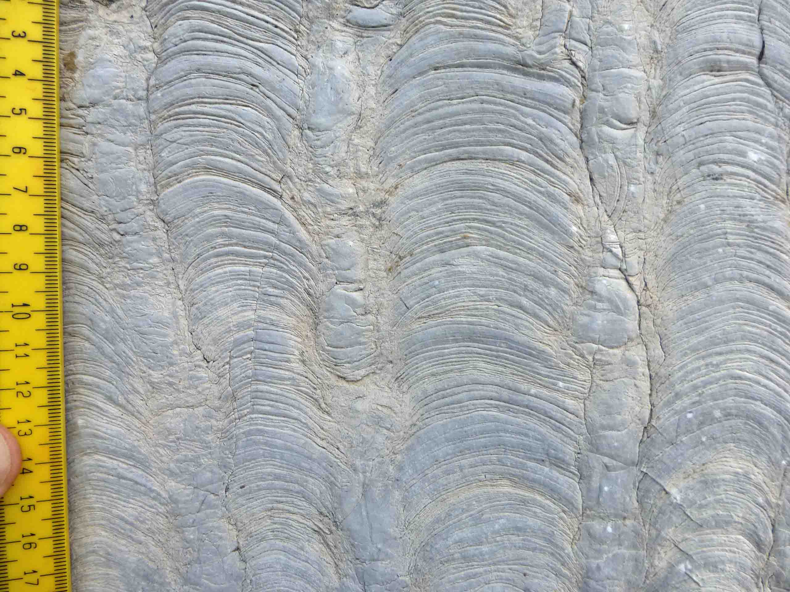
What are they, what are the differences between them and how they can improve your life?

1) WHAT IS IN THIS SECTION OF THE WEBSITE?

This section describes stromatolites and stromatoporoids, their similarities and differences and aims to clear up any misunderstandings about them. Stromatolites are often confused with stromatoporoids because they happen to have a similar name, and can look very similar when seen in rocky outcrops and even in polished rocks; but biologically they are not related. I will show you how to recognise these two structures, and how to distinguish between them. There are many places in the world (including the UK, where I am based) that have both of them, although not often together because they tend to have different requirements for growth. Nevertheless, there are cases where they occur in the same outcrops, so you can impress your friends (or simply reinforce their view that you are a bit weird) if you can distinguish stromatolites and stromatoporoids!

Stromatolites are very well known in the public domain; you don't have to look far to find descriptions and pictures of them, and there are many media descriptions of them in relation to evolution of life on Earth. They are constructed principally by bacteria and cyanobacteria, and are known to be amongst the oldest fossils on Earth, as well as being alive today in certain places. Fossil stromatolites grew mostly in the sea; living stromatolites are also in the sea, but there are examples which live in highly salty lakes and even in fresh water.

Stromatoporoids are sponges that grew a calcium carbonate skeleton and lived in normal marine conditions in shallow oceans. They have living representatives that occur in shady parts of the shallow sea floor, normally associated with coral reefs. However, in parts of the geological past, stromatoporoids were major reef constructors in their own right, and for tens of millions of years they occupied the same ecological position as modern coral reefs do. There is a lot of general information presented in this website in the section on Coral Reefs that you can access from the GENERAL INTEREST link on the Homepage. About 375 million years ago stromatoporoids were badly affected by mass extinction near the end of the Devonian Period of geological time, and since then have been much less important in shallow marine environments.

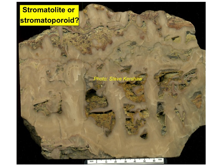
THE FOLLOWING PHOTOGRAPHS SHOW YOU HOW CONFUSING STROMATOLITES AND STROMATOPOROIDS CAN BE, WHEN YOU SEE THEM IN THE FIELD AND IN CUT PIECES OF ROCKS. TRY TO GUESS WHICH YOU THINK IS WHICH, THEN CHECK THE ANSWERS AT THE END OF THIS SECTION OF THE WEBSITE (you can cheat if you want to, but it won't help you when you find them for yourself).

Figure 1: one is a stromatoporoid, the other is a stromatolite.

Figure 2: one is a stromatoporoid, the other is a stromatolite.

Figure 3: Is this a stromatolite or a stromatoporoid?

Figure 4: Is this a stromatolite or a stromatoporoid?

Figure 5: Is this a stromatolite or a stromatoporoid?

Figure 6: Three of these are stromatolites, three are stromatoporoids, which is which?


2) SOME MORE BASICS ABOUT STROMATOLITES AND STROMATOPOROIDS

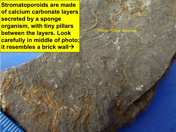
The reason for the similarity of the names of stromatolites and stromatoporoids is because they are similarly layered; the word stroma comes from Greek language, meaning a layer or mattress. Thus stromatolite means "layered rock", while stromatoporoid means "layered with porous structure". In most cases, stromatolites are composed of fine-grained finely-layered sediment which is cemented to preserve the layers. Stromatoporoids, in contrast, are calcium carbonate fossils, with an architecture that is highly variable, but is dominated by layers often called laminae and vertical structures often called pillars; some species of stromatoporoids are not dissimilar in appearance from the mortar between bricks in a brick wall (SEE PHOTOS). Thus stromatoporoids are equivalent to other fossils you might be familiar with (e.g. ammonites, trilobites) that have a hard skeleton which is preserved, while the soft tissues rot away. Stromatoporoids are therefore skeletal fossils, while stromatolites are mostly not, but there are some which are skeletal, and that makes the differences a little more complicated. I have not included skeletal stromatolites in this webpage, but if you are desperate to find out about them, then I could include them in a future update.

The oldest stromatolites are around 3,500 million years old, way back in the earlier part of the Precambrian time, in a part of the geological record called the Archaean Eon. Stromatolites are more abundant in the younger parts of the Precambrian time, the Proterozoic Eon. Most stromatolites are made of sediment compiled in layers, by the action of cyanobacteria and bacteria which colonised the sea floor, trapping sediment. It is commonly interpreted that the origin of oxygen in Earth's atmosphere is largely due to the photosynthesising action of stromatolites, over hundreds of millions of years. The Earth atmosphere was probably almost all carbon dioxide in its early history, as is still true for our two nearest neighbours, Mars and Venus, that lack life as we know it, Jim.

The oldest stromatoporoids, however, are much younger, because they are part of the events in later Earth history when skeletonised fossils became abundant. The earliest stromatoporoids might be late Cambrian Period, but are debated in relation to their biological position, because much depends on the interpretation of the nature of fossil structures that are not found as modern organisms. Nevertheless, definite stromatoporoids became abundant in the middle part of the Ordovician period, around 470 million years ago, and were the major reef-building fossils in this earlier episode of abundant skeletonised fossils, called the Palaeozoic Era. Stromatoporoids suffered extinction along with other fossils in the Late Devonian mass extinction event, in contrast to stromatolites, which are common after that extinction. Some researchers claim there is evidence that stromatolites (and other microbially-related structures) bloomed after extinction events, taking advantage of the abundant nutrients left available when large numbers of skeletonised creatures died out. The problem is that some of the major extinctions were not accompanied by stromatolites and their kind, so there is a lot of debate about that!


From the above paragraphs you can see there are major differences between these two fossil types, that are somewhat hidden by their superficial physical similarity; this is why they are worth knowing about.


ANSWERS TO FIGURES 1-6:

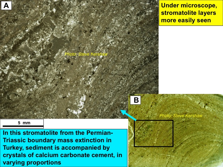
Fig. 1A: Stromatolite in southern Turkey from shallow marine environments directly after the end-Permian mass extinction event; Fig. 1B: Stromatoporoid from a Silurian shallow marine reef in Wenlock Edge, midlands of England. To be honest, from these photos, without prior knowledge of those sites, the two fossils are not distinguishable from these photos.

Fig. 2A: Stromatolite from a Silurian reef on the island of Gotland, Sweden; it is unusual to find them so well-developed in these reefs; Fig. 2B: Stromatoporoid from another Silurian reef, also on Gotland. In both cases, layering is well-displayed. Again, as in Fig. 1, it would hard to distinguish them in these photos, you need at least a hand lens, and maybe a microscope.

Fig. 3: Stromatoporoid from a Silurian reef on Gotland, Sweden. The rather flat structure and the small dome-shaped lumps on the upper surface near the top of the photo are more characteristic of stromatoporoids, but you could not prove it from this picture.

Fig. 4: A classic stromatolite, a piece of the Cotham Marble from the latest Triassic Period, cut in vertical section. This piece came from near Bristol, UK. The very thin multicoloured layers are very fine-grained sedimentary particles of calcium carbonate, and there are small sedimentary breaks shown by tiny erosion surfaces cutting the sediment layers. The odd-looking vertically orientated bubbly features are probably due to a change in growth structure caused by environmental change. This is certainly not a stromatoporoid. By the way, this rock is a limestone, not marble; marble is a metamorphic rock, metamorphism is a process of heat and pressure that destroys the original fabric of sedimentary rocks. In contrast this photo shows a beautiful sedimentary rock, with all its features preserved, and no indication of metamorphism.

Fig. 5: A stromatoporoid made of tall thin columns, linked together by lateral flanges. Stromatolites do not look like this. Lower Silurian, Gotland, Sweden.

Fig. 6: A, E and F are stromatoporoids, B,C and D are stromatolites. The only one you could be reasonably sure of is B, because of its very narrow columnar fabric. However, to distinguish them with certainty, you need a hand lens or a microscope.

For information, all the photos in this section of the website show limestone; all stromatoporoids were constructed from calcium carbonate (although in some cases become later silicified); nearly all stromatolites are made of layers of sedimentary material made of calcium carbonate, but there are some which were made from silica and even other materials, such as iron oxides. Just to make it more exciting, although most carbonate stromatolites are made of layers of sediment, some are made of crystallised calcium carbonate, called carbonate cement, for example Fig. 6B above, but you can’t tell from this magnification; finally some are made of a mixture of deposited particles of sediment and cement, and thus are hybrids (originally described by my good friend Robert Riding in a paper a few years ago). You will see some examples of this variation in the photos lower down.


TOOLS YOU NEED TO STUDY STROMATOPOROIDS AND STROMATOLITES; and some other information

You need a good hand lens of 10x magnification; you can use 20x (more expensive), but 10x is fine. With a 10x hand lens you can see the structure that makes up the fossil, and you can see whether the fossil is a stromatolite or a stromatoporoid. However, please note that using a hand lens to study a rocky surface will make you look nerd-ish, especially if you do it in public. There are numerous places in cities in the UK where polished facing stones on outsides (and insides) or buildings contain structures that have fine details needing a lens; thus if you use a hand lens to study them, either have lots of friends who are doing the same thing so that the general public steers well clear of you, or make sure you wear normal clothing; then if anyone bothers you, you can leave and blend into the crowd and not be noticed. PLEASE never damage polished rock surfaces on buildings; most fossils are in limestone, which is relatively soft (a knife would scratch it). They are for everybody's enjoyment, so leave them untouched.

If you want to go the whole hog and study these fossils in detail, you need a petrological microscope, and need to have rock thin sections (slices of rock so thin they are transparent, stuck to glass microscope slides); then you can see the details in all their magnificent glory. In this part of the website, and in other projects of this website, there are numerous photographs of rock thin sections taken through a petrological microscope, so you can appreciate the value of such tools. Making thin sections is not difficult, but you need to know how to go about it, and have the right equipment, and take the appropriate safety precautions. You also generally need permissions to collect samples, and it is essential to ensure you follow safety codes in the field.

SPECIAL NOTE: the best place to study stromatoporoids, that I know of, is the Exhibition Road entrance of the Natural History Museum in London; the entire entrance lobby is faced with the most beautiful layered stromatoporoids, in which their structure can be very clearly seen. Unfortunately, I don't know of any stromatolite facing stone in London, but there are lots of websites that illustrate stromatolites; there are even websites with "stromatolite" in the domain name! Please note that some websites claim that stromatolites have healing powers; however, they are really just pieces of limestone that have been created in layers, not a justification for such beliefs. Believe what you want to, of course, but if you decide to buy pieces from websites, you will be enriching yourself scientifically with some of the most beautiful geological material, but try not to pay too much. Most sites I have seen selling such material tend to charge too much, but even that is preferable to collecting your own, especially if you do not have polishing equipment, because stromatolites and stromatoporoids remain rather uninteresting lumps of limestone until they are polished.

If you find the information in this part of the website interesting, then you may want to look at the detailed coverage of some of my research into stromatoporoids and stromatolites in the GEOSCIENCE RESEARCHERS part of this website. You will see that some of the photographs shown in the pages here are repeated elsewhere in the website; this is deliberate, so that you can relate this section to others in the site.


3) A DESCRIPTION OF STROMATOLITES

Stromatolites are apparently simple structured, consisting of layers of sediment, but they vary. The following pictures are from a field visit organised by the China University of Petroleum (Beijing), associated with the first conference on Palaeogeography, Beijing, September 2013. The rocks are the Late Precambrian Upper Tieling Formation 1400 million years old, and the photos show you some of the variety of the stromatolites in this area. The stromatolite deposit is 100 m thick!!! Stromatolites are interpreted to have been largely responsible for the 21% oxygen of the atmosphere surrounding the Earth, and so a deposit 100 m thick of just stromatolites, is a small indicator of how abundant and important they were. Note that these pictures do not show all the variety of these Precambrian stromatolites, but after looking at the photos you should get a a useful understanding.

Figure 7: Upper left: view of a landscaped quarry where the stromatolites are preserved as part of a geopark. Upper right: a stone marking the presence of the stromatolites, written here in Chinese. Lower left: a view of columnar stromatolites in this site. Lower right: part of a description carved into a block of stromatolite next to one of the several sites in this area.

Figure 8a: Columnar stromatolites, with fine-grained sediment in between them that is not layered. There is a fragment of a stromatolite in between two columns in the big picture. Note that, in contrast, stromatoporoids rarely grew such columnar structures, so there is very little likelihood you will see stromatoporoids that look like this. Nevertheless, any good scientist would want to check for the layered structure that lacks the brick wall appearance of stromatoporoids, before being 100% certain of their identity.

Figure 8b: four pictures of variations in the stromatolite. Upper left: side view of laminated stromatolite. Upper right: a mixture of columnar and laminated forms. Lower two photos: along one particular layer, the stromatolite columns are bent and pressed against each other; this is rather odd, and they may have been affected by currents that forced them to grow in one orientation.

Figure 9a: a small cluster of small broken stromatolite columns that have fallen down in the gap between two large columns. The small columns thus seem to have broken along the curved growth surfaces, indicating that although they were reasonably solid, they could separate along the curved surfaces and fall into short lengths of column; thus they were not fully lithified (turned to stone) at the time they grew. Such features can be explained if the stromatolite layers were made of bacteria/cyanobacteria that trapped fine sediment, and was only later lithified.

Figure 9b: Close up view of some of the bent stromatolite columns, but at the top of the photo is a large jagged line that cuts through the stromatolites; the overlying stromatolites were compacted down onto the ones underneath. The jagged line is called a stylolite and is caused by the pressure of the overlying rocks when these deposits were buried in the Earth crust. Thus the stromatolites have suffered pressure solution, a very common feature of limestones, and is also illustrated in the Coral Reefs project on this website, accessed from the For Everybody page.

Figure 10a: Photos of columnar stromatolites in vertical and horizontal section, in a site near the top of the stromatolite deposit.

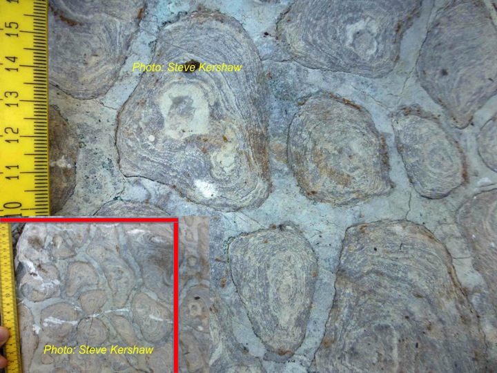
Figure 10b: Another view of some of the other stromatolites in the Upper Tieling Formation that shows the columns cut in horizontal cross section. The concentric rings are visible because the curved columnar structure is cut flat across, rather like the rings of an onion when cut through along a flat line.

Figure 11: three polished samples of vertically cut stromatolites, showing the beautiful smooth curving laminae of the successive growth layers. Note the brown and light grey unlaminated sediment between columns that filled the space after the stromatolites grew.

Figure 12: Field visits by geologists always have a group photo; here is the happy group of people who are smiling because of the amazing stromatolites! This particular field visit was sponsored and organised by the International Association of Palaeogeographers, based in Beijing, as part of the First International Palaeogeography Conference, Beijing, September 2013. With thanks to Prof Feng Zhengzhao.


The following photos are of stromatolites in thin section, taken down a microscope. These photos are from the stromatolites that grew after the end-Permian mass extinction, 250 million years ago, and are therefore much younger than the Precambrian stromatolites of the previous pictures.


Figure 13: Microscope thin section photos of the same specimen illustrated in Fig. 6D above, from the famous stromatolites that formed directly after the end-Permian extinction event, that killed 90% of all marine species. The stromatolites are part of a series of microbialite sedimentary rocks that replaced the ancient coral-sponge reefs that existed in the shallow marine environments before the extinction. These and other thin section pictures in this website were made by photographing a very thin slice of rock using transmitted light on a microscope. The black rectangle in photo B shows the area of photo A. Photo A lower right part is purely sedimentary stromatolite, but the left hand side has some tiny calcite (calcium carbonate) crystals, so this sample is a hybrid stromatolite.

Figure 14: More thin sections from stromatolites that formed after the end-Permian extinction. Photos A and B are sedimentary, Photo C is a mixture of sediment and cement, but Photo D is purely cement.

Figure 15: Another hybrid stromatolite.


3) A DESCRIPTION OF STROMATOPOROIDS

Stromatoporoids are beautifully layered structures made of calcite (calcium carbonate) skeletons. They started growing as layers that build up, layer by layer, like stromatolites do. However, stromatoporoids are sponges, filter feeders, that collected particulate organic debris floating in the water. The sponge tissue draws water in through pores in the surface and filters out the organic particles for food and absorbs oxygen from the water. Then the depleted water is collected together into larger tubes and expelled through centralised exit tubes; this is the reason why many of the photos below show rather beautiful root-like tubes that coalesce into a central point. These coalescing tube systems are called astrorhizae, and are distinctive characters of many sponges.

Figure 16: A broken piece of Devonian stromatoporoid from northern Spain; this sample was collected from an outcrop during a field trip in 1995 led by Sergio Rodriguez and Isabel Mendez-Bedia. You can see that natural weathering of the rock has accentuated the minor differences of resistance of the skeleton compared to the tiny spaces inside the structure, called gallery space. Thus the intricate structure can be seen in this photo. Consequently, you should look carefully for this structure if you are studying stromatoporoids for yourself.

Figure 17: A cut and polished piece of Devonian stromatoporoid, this time it is from the Anderdon Quarry, Ontario, eastern Canada, collected in 1983 on a field trip courtesy of Professors Al Fagerstrom and Carl Stock. The laminar-pillar structure and the banding can be seen clearly in this photo. The reasons for the banding are not clear; in some samples it is possible to show that the spacing of successive laminae varies, suggested by some researchers to be due to growth rate changes in the specimen, and some people interpret these as annual growth bands. This is not proved, but if it is true, can you work out the growth rate, and impress your friends?

Figure 18: This photo speaks for itself.

Figure 19: Microscope thin section photos of the stromatoporoid Petridiostroma, showing the laminae and pillar skeletal structure with gallery spaces between the skeletal elements. Compare this with the stromatolite microscope photos earlier on this page.

Figure 20: Cut and polished vertical sections of specimen of the stromatoporoid called Labechia, from Coates Quarry, Wenlock Edge, UK, in Upper Silurian rocks of the Wenlock Series. You can see the prominent thick pillars that characterise this stromatoporoid, and it shows that not all stromatoporoids are dominated by the laminae.

Figure 21: Vertical and horizontal thin sections of Labechia emphasising the pillars. The laminae do not really exist here, but the pillars are separated by curved plates, called cyst plates.

Figure 22: These two samples are in plan view, in which the astrorhizae are visible. Astrorhizae are visible only in plan view (also called tangential section). The left photo is a piece of stromatoporoid photographed showing its surface, so the astrorhizae are actually grooves in the surface. The right photo is a thin section, cut somewhere in the interior of the specimen. NOTE: these two photos are from different genera of stromatoporoid, in fact the left one is from the lower Wenlock, the right one from the middle Ludlow, both in the Silurian period. If you have time do look up on the internet these geological names and find out what ages they are; there are also some very useful smartphone apps that give you the geological timescale that you can carry in your pocket, and impress your friends.

Figure 23: Vertical and horizontal thin sections of the stromatoporoid Eostromatopora, from Wenlock Edge, Upper Silurian of UK. Note that the atrorhizae are visible in horizontal section (C), but not in the two vertical sections (A & B); the astrorhizae are there, but you cannot see them in vertical section. In photo A, the dark stuff upper left is the fine-grained sediment that was deposited on the sponge after it died. Also in A, the dark grey blob with a hole in it (looks a bit like the Cyclops, ha ha !!) that is stuck to the top surface of the stromatoporoid; this is a crinoid, a very common fossil in Silurian limestones.

Figure 24: Vertical and horizontal microscope sections of the stromatoporoid Plectostroma, showing prominent pillars connected by thin rods; thus, like Labechia, it does not have true laminae, yet the whole sample shows a layered structure because of growth variations throughout the development of the specimen.

Figure 25: Vertical and horizontal microscope thin sections of the stromatoporoids Syringostromella and Ecclimadictyon, showing the variations in the structure. Much Wenlock Limestone Formation, Upper Silurian, Wenlock Edge, UK.

Figure 26: Vertical and horizontal microscope thin section of stromatoporoid Densastroma; it shows a very dense skeletal structure, so that the laminae and pillars are almost impossible to see; however, you can clearly see the astrorhizae in (B). Photo A shows some other fossil organism is present within the stromatoporoid (the white ellipses), probably some kind of worm. Photo C is tremendously interesting for a different reason, as explained below.

Make a cup of tea or coffee and sit down before reading the following (you might also need a biscuit or piece of cake, try the cheesecake recipe described in the Dark Zone). There is a continuing controversy about the what mineral that stromatoporoids were originally made from; they are composed of calcium carbonate, but there are three different mineral forms of calcium carbonate that are commonly found in fossil organisms. All stromatoporoid fossils from the Palaeozoic Era are made of calcite (the most common form of calcium carbonate, but it is not clear what kind of calcium carbonate they were originally made from, because there are three different forms: aragonite, magnesium-rich calcite (called Hi-Mg calcite) and magnesium-poor calcite (called Lo-Mg calcite). These three forms of calcium carbonate are used by carbonate-secreting organisms and all three occur in fossils. But the problem is that stromatoporoids are always altered from their original mineralogy, so the question remains as to what was their original mineralogy. This is not a meaningless question; geoscientists have built oceanographic theories on the basis of the type of calcium carbonate found in limestones through geological time, and the original mineralogy of stromatoporoids plays a part, because they were so abundant. There is a way to test the original mineralogy of stromatoporoids, by comparing them with the minerals of other fossils found along with them, in the same samples, and often in the same thin sections. In Photo C the lower curved structure, that forms the surface upon which the stromatoporoid grew, is actually another fossil, it is a gastropod (snail). If you look carefully you can see that the dark grey sediment that filled the gastropod tube is actually directly in contact with the base of the stromatoporoid. This means that the gastropod shell is no longer present, it has been dissolved away and the stromatoporoid has compressed down onto the sedimentary infill of the gastropod. This kind of feature is very common in the geological record and occurs because the gastropod shell was made of aragonite; clearly it was less stable than the calcium carbonate of the stromatoporoid. Gastropods belong to the mollusc group of animals, that are typified by aragonite skeletons. Thus we can deduce that this stromatoporoid was NOT made of aragonite. If you get into the fossil literature you will find that some people claim stromatoporoids were made of aragonite. Because all stromatoporoids are recrystallised, but do not dissolve away, this is evidence that they were not originally aragonite, and that leaves Hi-Mg calcite and Lo-Mg calcite as the other two possibilities. However, it is unlikely that stromatoporoids were made of Lo-magnesium calcite, because that rarely becomes altered, so that leaves Hi-magnesium calcite as a reasonable alternative, yet proof is still not achieved. The problem is that the recrystallised fabric of stromatoporoids looks completely different from the fabric of fossils known to be of originally Hi-Mg calcite mineralogy. So it is a conundrum, that you can REALLY impress your friends with, or possibly send them to sleep. Certainly if you read anywhere that an author has claimed that stromatoporoids were aragonite, or were Hi-Mg calcite, you have here the evidence that makes it difficult to identify the original mineralogy. You might like to download a paper that gives you some more background: http://www.journalofpalaeogeography.org/EN/volumn/volumn_1138.shtml.

OK, you can relax now, no more about stromatoporoid mineralogy! (How was the cheesecake?)

Figure 27: Vertical section of stromatoporoid called Parallelopora; this specimen contains lots of tubes of other fossils, probably some kind of worm, that grew symbiotically along with the stromatoporoid. The co-growth of the tubes helps us to understand about the growth of the stromatoporoid on the small scale; the different coloured arrows point to different relationships between the stromatoporoid and its symbiotic tube animals. Yellow arrows show tubes where the laminae abut against the tube with no deflection; blue arrows show tubes where the laminae are deflected in contact with the tube. The green and red arrows point to spiral tubes that are cut through the spiral; notice the lower tubes in each cluster are smaller diameter, and the higher tubes increase in diameter, they are therefore increasing diameter while spiralling upwards, like an ice cream cone if it was twisted into a spiral. The red arrowed tube seems to begin at a prominent break in the growth of the stromatoporoid, shown by the change in grey colour of the stromatoporoid skeleton. The green arrowed tube seems to begin above a prominent break. These two different spiral tube start points might reflect the ability of the spiral tube animal to get a hold on the surface of the stromatoporoid. In all the cases of these symbiotic tubes, the stromatoporoid seems to accommodate its host, and leave the impression that the stromatoporoid did not suffer as a result of these guests. Whether or not the stromatoporoid benefited from their presence is open to debate, but there seems to be no obvious advantage; that would lead to the interpretation that they tolerated the symbiotic tubes without an ability to kill them.

Figure 28: These pictures show a coral attached to the upper surface of the stromatoporoid. Look closely at photo B; the top of the stromatoporoid shows an irregular shape, and suggests that it was eroded before the coral attached. Such an arrangement can be interpreted that the stromatoporoid died, was eroded and then the coral attached. If so, the coral was simply using the stromatoporoid as a hard place to attach, to give it a good base for growth.

Figure 29: This is my favourite: a horizontal thin section of a stromatoporoid that has not one but TWO corals that lived and grew with the stromatoporoid. Thus there are three fossils here, a remarkable and very beautiful result; this specimen shows the ability of stromatoporoids to accommodate their guests. This stromatoporoid is called Petridiostroma convictum, from the Upper Silurian of Gotland, Sweden; it is also found in Estonia, 250 km WNW of Gotland. Every single specimen of this species that I have seen (hundreds of specimens) has at least the small coral symbiotically inside it, and very often has both corals, a truly remarkable phenomenon.

Figure 30: A summary synthesis diagram in 3D that shows a range of the major appearances of stromatoporoid skeletal structure (this is an unpublished diagram from my PhD thesis, which was written in 1979; it shows that even 35 years later, there is still something useful to come from my PhD!!!).

Figure 31: In case you have not realised it, this is a joke; there is no stromatoporoid that looks like this, and there is no stromatoporoid called Distressostroma perplexa, but if you spend months studying them under the microscope, this is what they look like at the beginning!!


4) THE ENVIRONMENTAL SETTING OF STROMATOLITES AND STROMATOPOROIDS

The following photos show the kinds of situations in which stromatolites and stromatoporoids may be found. The pictures show some modern settings followed by rocky outcrops of ancient reefs. There are also a couple of comparative photos at the end that show some more interesting things.


Figure 32: These pictures show the environmental setting of stromatolites and stromatoporoids. Photo A shows the modern Great Barrier Reef, made of corals; this is a model for the ancient coral-stromatoporoid reef from the Silurian, which is shown in B. C is the modern stromatolite formation of Shark Bay, Western Australia, often considered the model for the Precambrian stromatolites seen in Figures 7-12. D is marked to show that the ancient Silurian reef is really quite small; the vegetation below the letter D is in a position outside the reef, showing the reef is only a few metres across. Nevertheless, some ancient stromatoporoid reefs can be gigantic, many hundreds of metres across.

Figure 33: More photos of the small Silurian reefs, containing corals and stromatoporoids. A very interesting aspect of these reefs is that their margins are very sharp, which is rather odd, because you would expect the reefs to have debris falling into the surrounding sediment, but there is very little; why that is so, is explored in the next two figures.

Figure 34: Photo A shows the margin of a Silurian reef from England, with two enlargement photos taken under the microscope, B & C, from just outside the reef (B) and just inside the reef (C). The microscope pictures show a strong difference between the reef rock of C and the bedded sediment outside the reef, in B. This has been interpreted to mean that the reef margin was solidly cemented and the sediment around the reef was deposited against the already cemented reef rock. The interpretation of myself and my colleagues is that this is explained by microbial cementation making the reef solid, but the sediment deposited against the reef is made of uncemented debris, that becomes rock much later. The next photo develops this idea.

Figure 35: This is a microscope view of a vertical section of a reef rock from the Upper Silurian of Gotland; the rock consists of two main components. The light-coloured swirly grey material is interpreted as microbial cemented solid carbonate; this is called LEIOLITE, and is simply another type of microbial rock. In contrast, the dark material is called WACKESTONE, which is simply one type of fine-grained sediment with shelly fragments embedded in it. The wackestone is deposited amongst an open framework of solid rock of the leiolite. There are no stromatoporoids or stromatolites in this photo, but it shows that they were only one component of these ancient reefs; the growth of microbial structures, which can include these strange leiolites, are integrated components that help to build the ancient reef. Ancient reefs are complicated and very fascinating.

Figure 36: This picture is included as an illustration of another type of microbial structure called THROMBOLITE, because it looks like a clotted structure. Thrombolites are very common and abundant in ancient reefs, and are as important as STROMATOLITES. Please take a look at the section on Mass Extinctions on another page in the For Everybody section of this website, and you will see more photos of thrombolites and stromatolites. There are more in the Atlas of Microbialites After the End-Permian Extinction, accessible in the GEOSCIENCE RESEARCH section.

Figure 37: To finish Section 3, I added a rather nice photo of a fossil which has exercised the excellent minds of many geologists. This fossil is called Solenopora, it is a layered structure, and looks like a stromatolite; but it is composed of millions of tiny tubes, not visible in this photo, and people have puzzled about whether this fossil is a stromatolite, with some thinking it might even be a sponge!! Such are the peculiarities of studying fossils. This specimen also has another piece of magic; its beautiful pink colour. This specimen, from west England, is Middle Jurassic in age and is from a rock called the Beetroot Stone, because of its colour. The surrounding sedimentary rock does not have the pink colour, so the implication is that the pink reflects some original colour remnant of the living organism. One interpretation is that the colour is caused by the presence of boron attached to organic chemicals in the once-living organism. Colour in fossils is not common, and opens a window to help us realise that the organisms we now see as rather dull grey, yellow, brown etc. colours, were originally likely brightly coloured creatures. We have no clear idea of the original colour of most stromatolites and stromatoporoids, but it is interesting to consider the possibility that they had a kaleidoscope of colours that are seen in modern reefs.


4) WHAT ARE THE WIDER IMPLICATIONS OF THE EXISTENCE OF FOSSILISED STROMATOLITES AND STROMATOPOROIDS?

Stromatolites and stromatoporoids are very abundant fossils in the time periods in which they occur in the geological record. Stromatolites date from 3,500 million years ago, and likely formed the oxygen of the atmosphere that we breathe, because they were photosynthetic. Stromatoporoids are younger (and as animals there is no evidence that they were photosynthetic), but there were vast numbers of them growing in the sea for about 100 million years in the Ordovician, Silurian and Devonian Periods from about 470 to 370 million years ago. As living creatures they both did one thing very well: they took carbonate from the seawater, combined it with calcium in the sea, to make calcium carbonate to form their structures on the sea floor. Where did the carbonate come from? The only viable source is the atmosphere as carbon dioxide that dissolved in the ocean and was transformed into carbonate by a well-established chemical process.

It is modelled that the early Earth had an atmosphere much richer in carbon dioxide than now, indeed there is so little carbon dioxide left that it is almost an insignificant component of the atmosphere if it did not have such powerful greenhouse properties that most people consider to be a cause of global warming. Thus stromatolites especially, and stromatoporoids to a lesser extent, were responsible for the removal of large amounts of carbon dioxide from the atmosphere, locking it up on the sea floor, and ultimately burying it in the surface layers of the Earth crust as the common rock LIMESTONE. You can see limestone all over the world. In most places in the world, you cannot go far without encountering limestone, of all different geological ages.

Thus I hope you can appreciate that these fossils are not just rather beautiful curiosities; they played a significant role in climate change through geological time. Without the oxygen in the air, higher animals such as humans would not be able to exist, because we need to oxygen to respire stored organic energy (food) to allow us to undertake the highly energetic occupation of the planet that we do. Therefore we should offer thanks to these lowly creatures, and go on field trips to study them and appreciate their place in the history of the Earth surface.


Click here to return to the GENERAL INTEREST PROJECTS header page

Click here to return to the HOMEPAGE