THE FIRST DISCOVERY OF SPICULES IN PALAEOZOIC STROMATOPOROIDS
A Devonian stromatoporoid from southern Belgium shows that spicules did exist in Palaeozoic stromatoporoids
1) WHAT IS IN THIS SECTION OF THE WEBSITE?
The information in this project is based on a recently published paper: Da Silva, A-C., Kershaw, S., Boulvain, F., Hubert, B., Mistiaen, B, R., Reynolds, A. and Reitner, J. 2014. Indigenous demosponge spicules in a Late Devonian stromatoporoid basal skeleton from the Frasnian of Belgium. Lethaia, DOI: 10.1111/let.12064. Lethaia is a major paleontological journal publishing high quality international peer-review work. The header page of this paper is given in Fig. 1.
The sample described here was discovered by Anne-Christine Da Silva.
The full names of the authors of this paper are provided, with addresses and emails, after the abstract below.
Figure 1. Screenshot of the header page for this paper, in the paleontological journal Lethaia.
ABSTRACT OF THE PAPER: This paper records the first example of a demosponge spicule framework in a single specimen of a Devonian stromatoporoid from the Frasnian of southern Belgium. The small sample (2.5 x 2 cm) is a component in a brecciated carbonate from a carbonate mound in La Boverie Quarry 30 km east of Dinant. Because of the small size of the sample, generic identification is not confirmed, but the stromatoporoid basal skeleton is similar to the genus Stromatopora. The spicules are arranged in the calcified skeleton, but not in the gallery space, and are recrystallized as multi-crystalline calcite. The spicules fall into two size ranges: 10 to 20 microns diameter and 500 to 2000 microns long for the large ones and between 5 to 15 microns diameter and 50 to 100 microns length for the small ones. In tangential section, the spicules are circular, they have a simple structure, and no axial canal has been preserved. The large spicules are always monaxons, straight or slightly curved styles or strongyles. The spicules most closely resemble halichondrid/axinellid demosponge spicules and are important rare evidence of the existence of spicules in Palaeozoic stromatoporoids, reinforcing the interpretation that stromatoporoids were sponges. The basal skeleton may have had an aragonitic spherulitic mineralogy. Furthermore, the spicules indicate that this stromatoporoid sample is a demosponge.
NAMES AND ADDRESSES OF THE AUTHORS:
Anne-Christine Da Silva [ac.dasilva@ulg.ac.be], and Fr�ed�eric Boulvain [fboulvain@ulg.ac.be], Sedimentary Petrology, Liege University, Boulevard du rectorat, 15, B20, Sart Tilman, 4000 Liege, Belgium; Stephen Kershaw [Stephen.Kershaw@brunel.ac.uk], Institute for the Environment, Brunel University, Kingston Lane, Uxbridge, UK; Benoit L. M. Hubert [benoith@icl-lille.fr], Bruno Mistiaen [bruno.mistiaen@isa-lille.fr], Laboratoire de Pal�eontologie stratigraphique, ISA Groupe, FLST, G�eosyst�emes UMR 8217 du CNRS, 41 rue du Port F-59016 Lille Cedex, France; Alan Reynolds [alan.reynolds@brunel.ac.uk], Experimental Techniques Centre, Brunel University, Kingston Lane Uxbridge, UK; Joachim Reitner [jreitne@gwdg.de], Department of Geobiology, University of Goettingen, Goldschmidtstr. 3, 37077 G€ottingen, Germany.
Manuscript received on 03/07/2013; manuscript accepted on 07/11/2013.
This paper is a contribution to the International Geoscience Programme projects IGCP591, 596 and 580.
Figure 2. Logos of the three projects to which this paper is a contribution.
1. INTRODUCTION
|
Stromatoporoids are calcified sponges, abundant in the Palaeozoic Era, during the ca. 100 million years between the middle Ordovician and Late Devonian Periods, ca. 470 - 370 Ma. They suffered in the Late Devonian extinction (the Frasnian/Fammenian event [F/F]) and almost completely disappeared from the rock record at the end of the Devonian Period (see Figs. 3 and 4). Upper Palaeozoic stromatoporoids are rare, but Mesozoic stromatoporoids are abundant and contain spicules, in contrast to Palaeozoic ones.
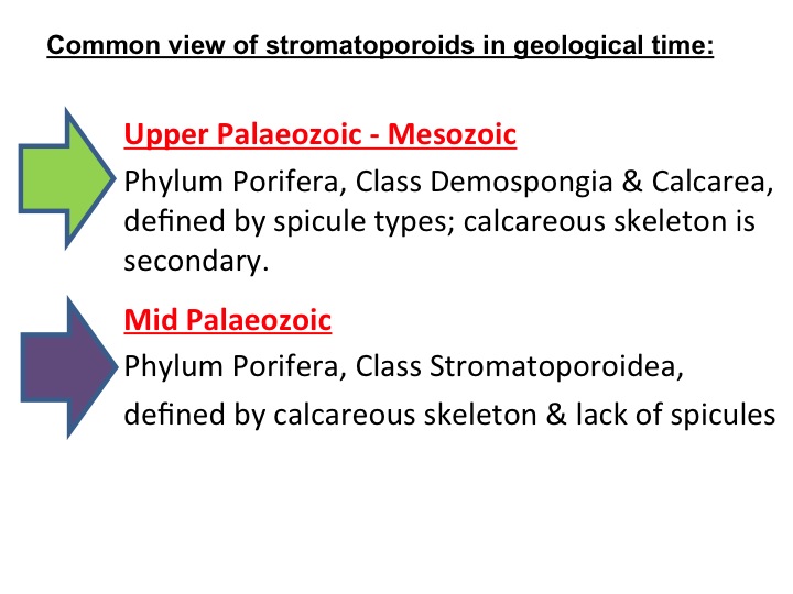
Figure 3. Simple graphic of key features of mid-Palaeozoic vs Upper Palaeozoic to Mesozoic stromatoporoids.
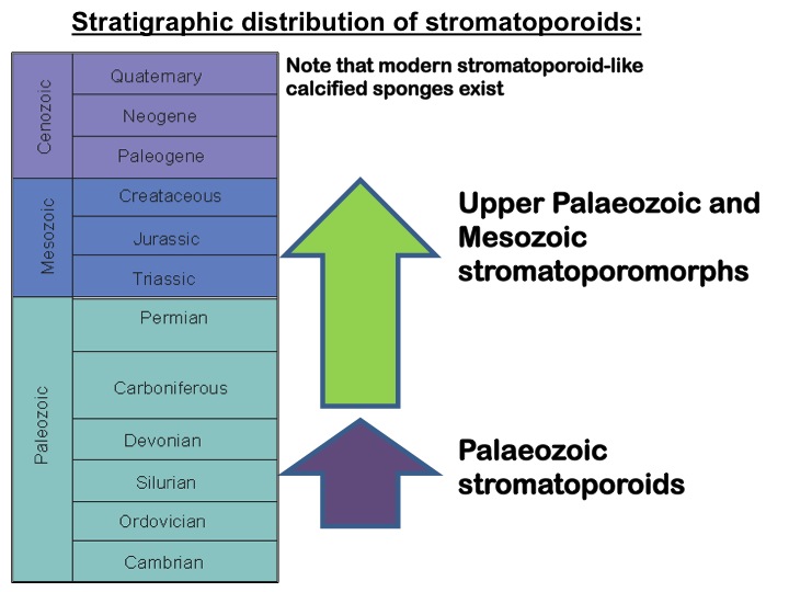
Figure 4. Stratigraphic ranges of the mid-Palaeozoic vs Upper Palaeozoic to Mesozoic stromatoporoid types.
Until now there has been only one description of spicules in Palaeozoic stromatoporoids (Wood et al. 1989), and that was in Carboniferous material, a long time after the Late Devonian decline of the major period of stromatoporoid development. This study reports the discovery of spicules in a Devonian stromatoporoid from the period before their major decline at the Frasnian-Famennian extinction event. For information about extinction events in Earth history, please see the Mass Extinctions project in the GENERAL INTEREST section of this website; the F-F extinction is one of the Big Five events.
The Frasnian Epoch is part of the Late Devonian Period, and was a time of major stromatoporoid reef development globally, including the reef sequences that contain the sample described here. In Southern Belgium, Frasnian reefs are abundant in the Ardennes region (see Fig. 5), where stromatoporoids are very abundant.
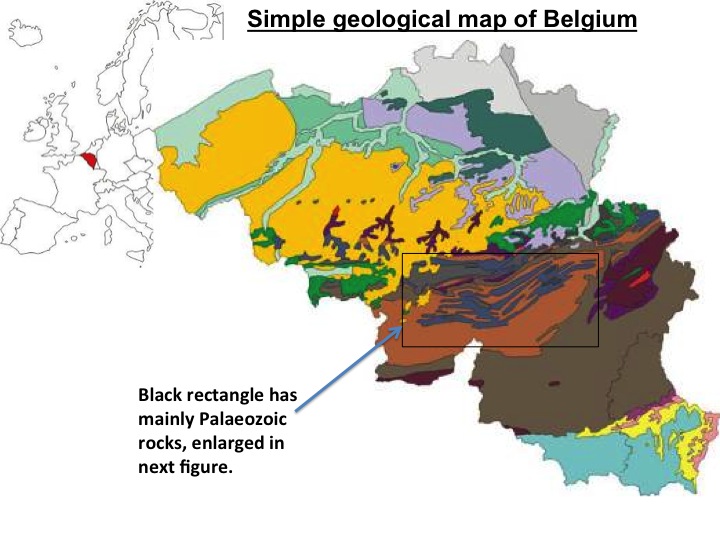
Figure 5: Location of Belgium and simple geological map of Belgium showing the Dinant synclinorium comprising Palaeozoic rocks of the study area.
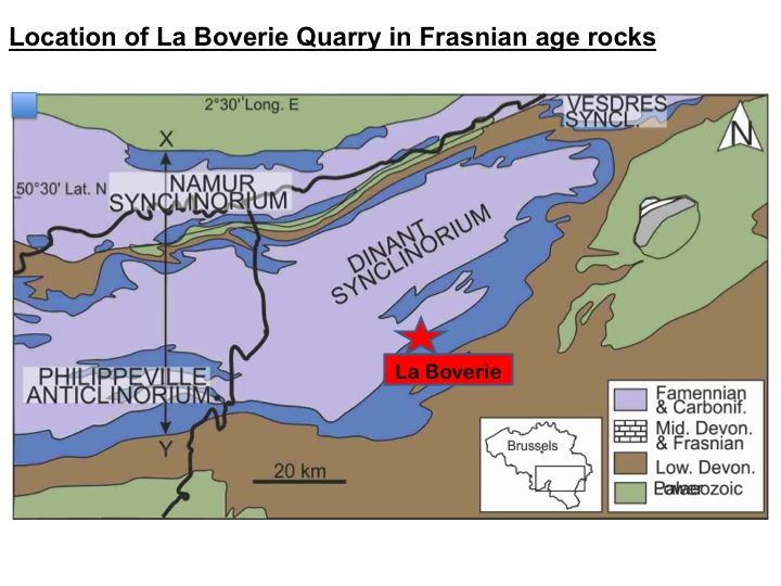
Figure 6: Simple geological map of the Dinant synclinorium, showing the location of the stromatoporoid found in this study, at La Boverie.
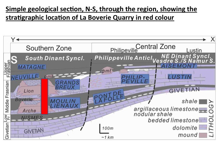
Figure 7: Simple N-S cross section through the Dinant synclinorium, showing the location and stratigraphic position of the stromatoporoid found in this study, in the Middle Frasnian Series, at La Boverie.
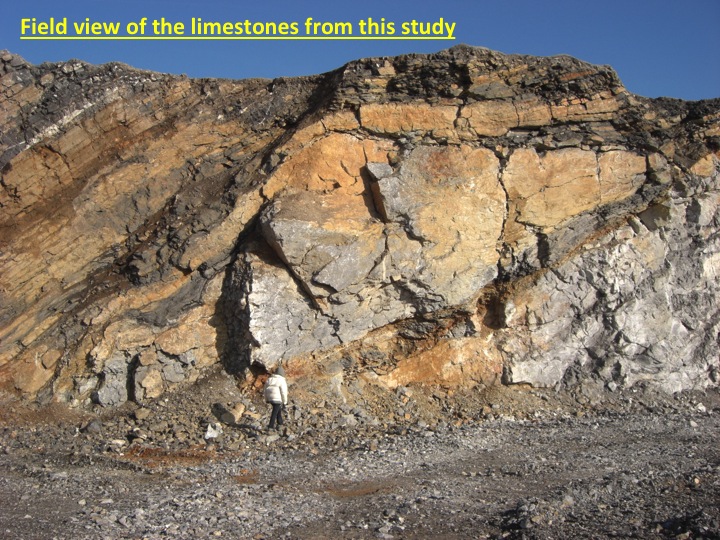
Figure 8: Field photograph of the La Boverie Quarry showing the tilted Middle Devonian limestones in which the stromatoporoid was found.
The Frasnian Series in southern Belgium is characterized by a succession of four mud mounds, which are in stratigraphical order: the Arche, La Boverie, Lion and Petit Mont mounds (Fig. 9; see also Boulvain & Coen-Aubert 2006).
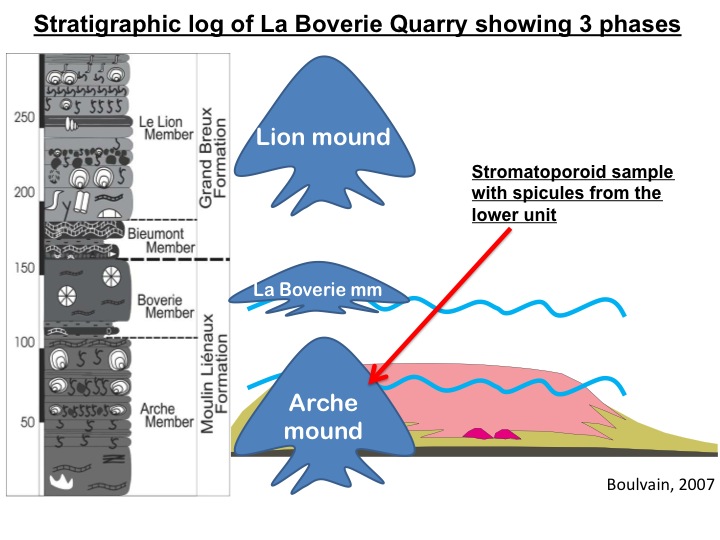
Figure 9: Stratigraphic log of the La Boverie Quarry, and schematic appearance of the three mounds occurring through the history of the quarry. The stromatoporoid sample came from the lowermost mound, the Arche mound.
The series of build-ups, including the Arche, La Boverie and Lion mounds, exposed in the quarry is nearly 300-m thick (Fig. 9). In the Central and Northern parts of the Frasnian Belgian platform (Fig. 7) biostromal and lagoonal facies dominate and are also rich in stromatoporoids (Da Silva et al. 2011b). The specimen described in this study comes from the middle part of the Arche mound (lower part of the Middle Frasnian), from a brecciated unit containing centimetre to decimetre-sized broken pieces of stromatoporoids and tabulate corals (Fig. 10).
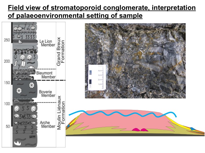
Figure 10: Field photo of the conglomerate and its position in a low stand episode of the Arche mound. The occurrence of this brecciated level is interpreted as related to a lowering of the sea level, leading to a reworking on the top of the mound and occurrence of these brecciated beds on the flank of the mound (Boulvain 2007). Thus, the horizon is interpreted to be lateral to the main mound body (Da Silva et al. 2010, 2011a).
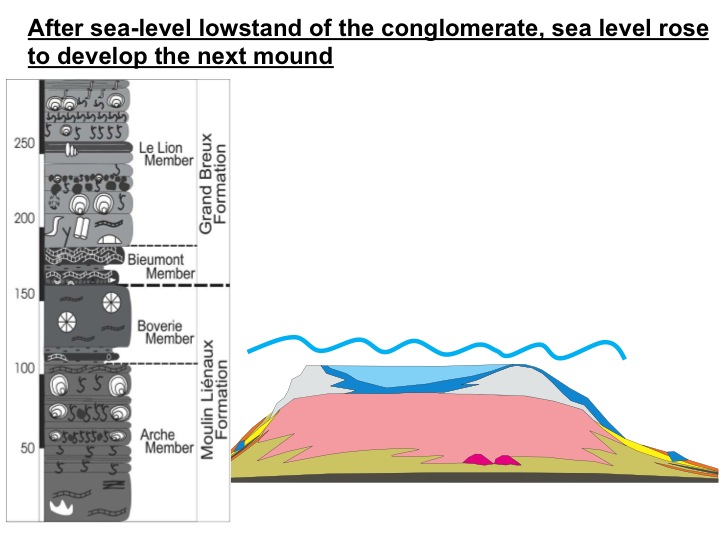
Figure 11: After the low stand that deposited the conglomerate containing the speculated stromatoporoid, sea level rose for deposition of the next mound.
The spicule-bearing sample turned up in a thin section and was a chance discovery; spicules in stromatoporoids cannot be seen in the hand specimen, and thus was visible only under a transmitting microscope.

Figure 12: Polished piece of rock showing the fragment LBv 54 2 x 2.5 cm, that has the spicules. Its stratigraphic location is show by the arrow on the left column. The stromatoporoid sample is surrounded by dolomitic and sparitic cements. Four thin sections were made (one tangential, one longitudinal
and two oblique sections). The samples were examined under a normal light microscope, scanning
electron microscope and cathodoluminescence (CL), the last offering the best images. The specimen, is held in Li�ege University (Belgium). Stromatoporoid terminology comes from the Treatise Online of Invertebrate Palaeontology (Webby 2010).
2. DETAILS OF THE SPICULES
|
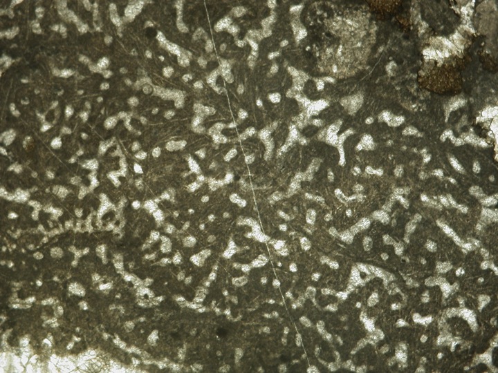
The following photos, Figs. 13 - 16, show increasing enlargement views of the sample in thin section in transmitted light, to show the spicules.
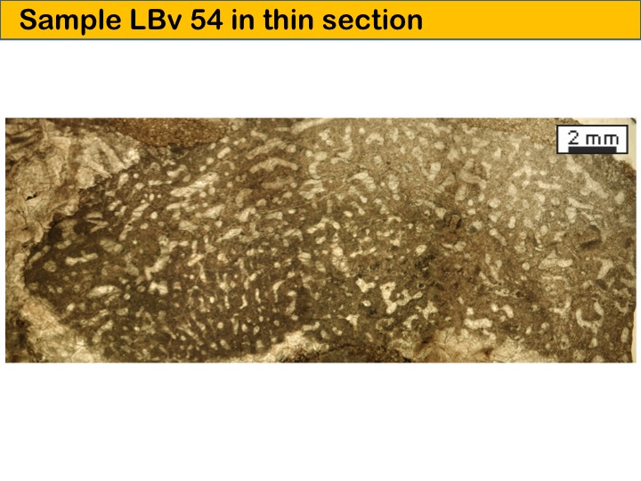
Figure 13: Thin section photograph of the entire fragment.
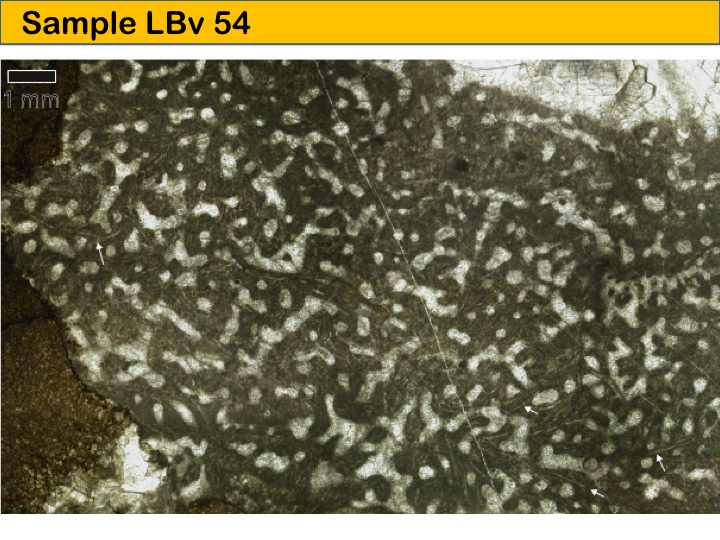
Figure 14: Enlargement, where spicules are visible as faint white lines in the stromatoporoid skeleton.
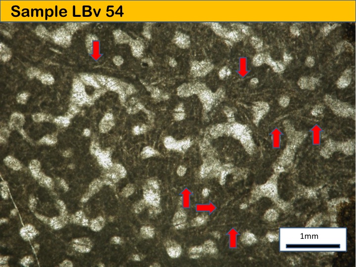
Figure 15: Enlargement, spicules much clearer in this picture.
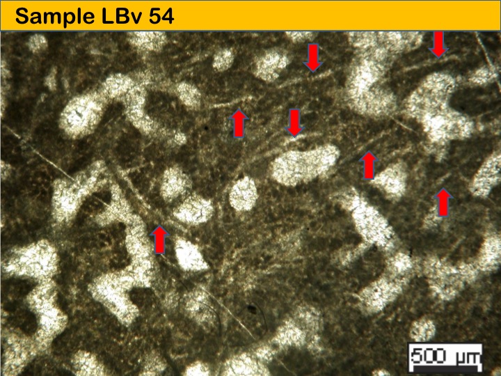
Figure 16: Enlargement, shows spicule morphology well and confirms that they occur only in the stromatoporoid skeleton, not passing into the gallery space.
One thin section was investigated by field emission SEM, using a LEO 1530 Gemini (Zeiss) instrument at 3.8 kV. The sample was polished prior to etching with 5% EDTA solution for 5 s to investigate micritic fabrics and blocky sparitic cements. Energy dispersive X-ray spectrometry (Oxford Instruments EDX) was performed on Au-coated samples using the same instrument operated at 15 kV. The results are shown in Fig. 17.
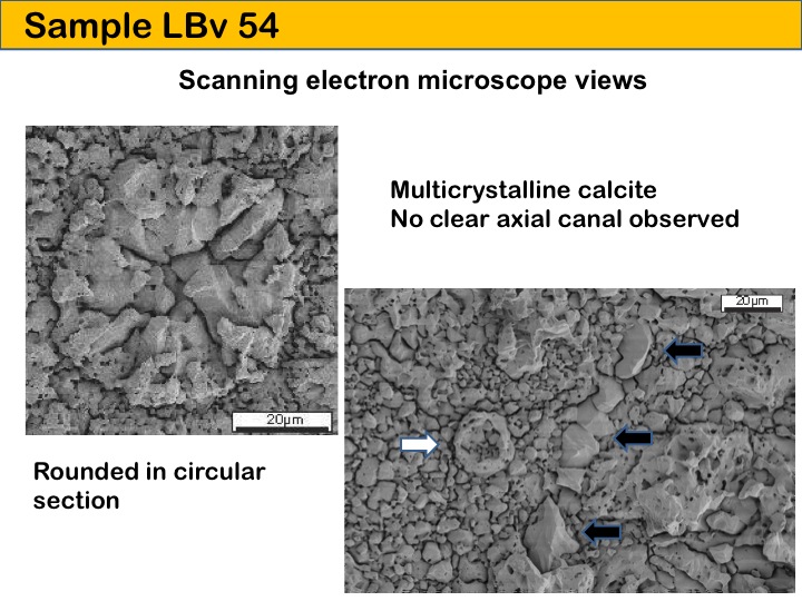
Figure 17: SEM photos showing the solid spicules, composed of multiple crystals with no central canal.
Cathodoluminescence investigations were carried out with a Citl 8200 MK3A cold cathode mounted on a Zeiss Axiolab microscope. Micrographs were recorded at 15 kV voltage using a cooled SPOTCCD camera. See Fig. 18. All facilities are hosted in the Geobiology laboratory at the University of Goettingen where the work was carried out.
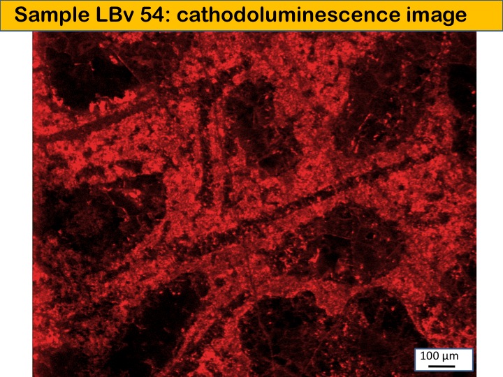
Figure 18: Cathodoluminescence photograph of non-luminescent spicules (dark lines) in the speckled red-orange colour skeletal structure of the stromatoporoid skeleton.
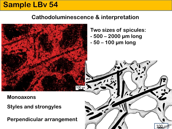
Figure 19: CL view of the stromatoporoid duplicated from Fig. 18, together with a reconstruction drawing of the spicules and stromatoporoid skeleton.
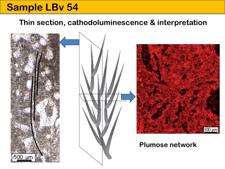
Figure 20: Normal light and CL views of the stromatoporoid and spicules in different orientations, together with a reconstruction drawing. This figure shows the three-dimensional view of the spicule arrangement.
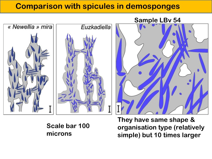
Figure 21: Comparative drawing showing the spicules of the specimen in this study (right hand image) against the Carboniferous specimen Newellia mira and the Mesozoic sponge Euzkadiella.
3. CONCLUSION
|
The photographs in this section of the website are a simple illustration of the first mid-Palaeozoic stromatoporoid to have verified spicules. It therefore reinforces the view that stromatoporoids were sponges. Furthermore this sample is a member of the demosponge group, based on the spicules. The sample therefore shows the possibility that the calcareous-based skeleton of stromatoporoids, while crucial for taxonomic identification at lower levels, may not be so reliable for higher level groupings.
Please see the published paper for a full discussion of this sample and its implications for the study of Palaeozoic stromatoporoids.

Figure 22. Reconstruction drawing of stromatoporoid skeleton and spicules, drawn by technical artists in Goettingen, whose work is gratefully acknowledged.
Click here to return to the GEOSCIENCE RESEARCH PROJECTS header page
Click here to return to the HOMEPAGE
| | |