Kershaw, Wood and Guo (2006) published a short paper in GFF on the relationship between stromatoporoids and their substrates. The issue is that Devonian stromatoporoids commonly display primary cavities beneath their skeletons, but this is poorly recognised in Silurian stromatoporoids. What are the reasons for this difference? The possibilities are:
1. Primary cavities are rare beneath Silurian stromatoporoids because they did not form such cavities easily. Thus stromatoporoids had the ability to grow directly on muddy substrates on which they commonly occur. That is in contrast to modern calcified sponges, which actively avoid fine-grained sediment.
2.Primary cavities beneath Silurian stromatoporoids may be common but simply have not been recognised because Devonian reefs contain much more precipitated calcite cement, that filled cavities and preserved them. Thus in the Silurian, cavities were closed by compaction prior to lithification, and are not preserved sufficiently for their importance to be demonstrated.
|
Assessment of the importance of primary cavities is a potentially valuable aspect of the study of Silurian and Devonian reefs. If stromatoporoids had capabilities to deal with fine-grained sediment in the Silurian, then perhaps one of the reasons why Devonian stromatoporoids developed into the peak of their occurrence is because of a change to form cavities, as an escape method to limit contact of the growing surface with muddy sediment.
This presentation was created to expand the study by Kershaw, Wood and Guo (2006) to examine substrate features of stromatoporoids, but includes tabulate corals because they have many growth features in common with stromatoporoids. The GFF paper was necessarily limited to a small number of pages, so this website expands the study with illustration of more material. >>
|
Thus the purpose of this section of the website is to illustrate fossils, to make available to the research community some detailed images that may be compared with other samples.
If you have any comments on any of this material, please email me at:
Stephen.kershaw@brunel.ac.uk
Reference: Kershaw, S., Wood, R.A. and Guo, L. 2006. Stromatoporoid response to muddy substrates in Silurian limestones. GFF, 128, 131-138.
|
The following images show the nature of contacts between stromatoporoids and their substrates. Further down this page I develop the ideas and present some material for which there is no single interpretation.

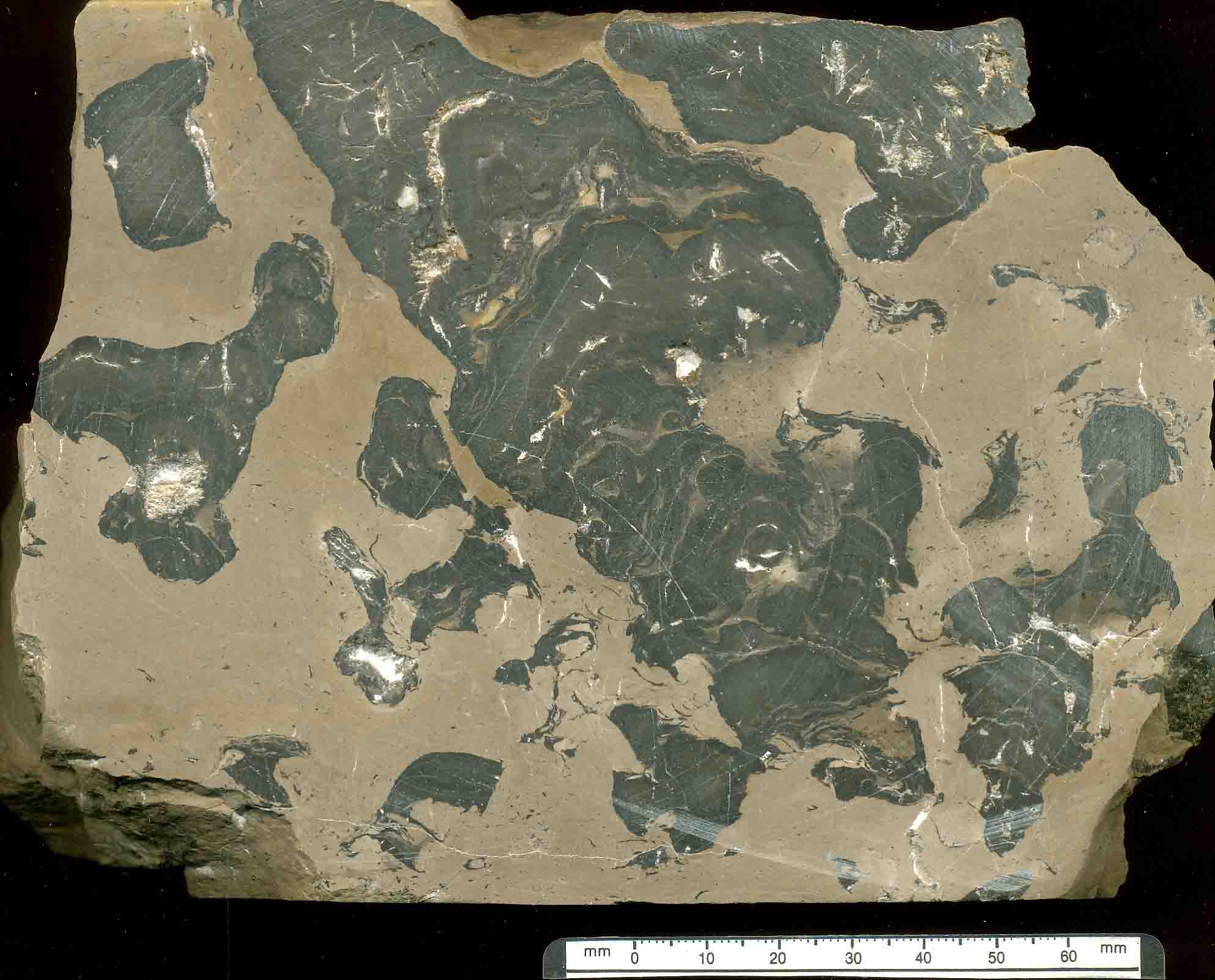
Figure 1.1. Vertical section of slab of Densastroma pexisum from Visby Formation, Lower Wenlock, Gotland, Sweden.

Figure 1.2. Vertical section of Pachystroma hesslandi from Visby Formation, Lower Wenlock, Gotland, Sweden.

Figure 1.3. Vertical section of Densastroma pexisum from Visby Formation, Lower Wenlock, Gotland, Sweden. Note the ragged margins.

Figure 1.4. Vertical section of thin section of Petridiostroma linnarssoni from Kneippbyn site, Visby Formation, Lower Wenlock, Gotland, Sweden. Note the response to sedimentation.

Figure 1.5. Basal surface of stromatoporoid from Visby Formation, Lower Wenlock, Gotland, Sweden. Note the encrusters on the base.

Figure 1.6. Basal surface of modern scleractinian coral Diploria, showing encrusters on base. Encrusters grew in a primary cavity formed by the overhanging of the coral from its central attachment area.

Figure. 1.7. Diagram showing views of interpretation of substrate relationships in stromatoporoids, from Kershaw (1998), The applications of stromatoporoid palaeobiology in palaeoenvironmental analysis. Palaeontology, 41, 509-544. Reproduced with kind permission of the Palaeontological Association.

Figure 1.8A. Vertical thin section of stromatoporoid from Upper Devonian of Belgium, showing primary cavity. Enlargement of box area shown in Figure 1.8B.

Figure 1.8B.
2) PRESSURE SOLUTION
|
A key problem in study of stromatoporoid palaeobiology is the common occurrence of pressure solution around the margins of stromatoporoids, especially in reef structures. >>
|
The photographs on this page illustrate the effects of pressure solution, showing stylolites in stromatoporoid margins from the Devonian reef at Long Quarry Point, Devon, England.
|

Figure 2.1. Vertical section of stromatoporoid showing the dome-shaped morphology, with ragged margins. Especially note the dark red-brown rims to the stromatoporoid, which are the stylolites, shown in enlargements in Fig.2.2.

Figure 2.2. Detail from stromatoporoid shown in Figure 2.1.

Figure 2.3. Another stromatoporoid showing stylolites at margins.

Figure 2.4A. Enlargement of Figure 2.3.

Figure 2.4B. Enlargement of Figure 2.3.
|
Now follows a series of pages of mostly Silurian stromatoporoids and corals from limestones from famous sites: Wenlock Edge, England; Gotland, Sweden; Anticosti Island, Canada. >>
|
Modern corals are illustrated for comparisons.
|
3A) EXAMPLES OF STROMATOPOROID-SUBSTRATE, AND CORAL-SUBSTRATE, RELATIONSHIPS
This section presents a range of examples of relationships between stromatoporoids and their substrates, and corals and their substrates. The purpose is to display a wide range of situations of relationships and to present interpretations and problematic cases. The material comes from:
Silurian of Gotland, Sweden;
Silurian of Wenlock Edge, UK;
Silurian of Anticosti Island, Canada.
STROMATOPOROID EXAMPLES

Figure 3A.1. Field setting of stromatoporoids and corals from the Visby Formation, Lower Wenlock, Gotland, Sweden. The facies is interbedded packstone-wackestone, and calcareous clays, of open shelf above storm wave base.

Figure 3A.2. Vertical thin section of Densastroma pexisum that grew on a dead orthoconic nautiloid. Presumably the stromatoporoid used the dead nautiloid as a small area of solid substrate to begin growth. However the right-hand edge of the stromatoporoid may have grown out into water, and then backfilled. It is not certain that this part of the sponge grew in direct contact with the sediment. However, on the left side, part of the stromatoporoid is on muddy sediment between the nautiloid and stromatoporoid, suggesting growth directly on the sediment. See next two figures in enlargement.


Figure 3A.3 & 4. Enlargements of Figure 3A.2.

Figure 3A.5. Complex growth of stromatoporoids, tabulate and rugose corals. The lower stromatoporoid (Densastroma pexisum) seems to have grown on an uneven substrate, possibly partially lithified. Sedimentation may have caused its death. Solitary rugose corals encrusted its surface, and there was further sedimentation that may have killed the corals. The upper stromatoporoid (Petridiostroma simplex) and a tabulate grew on a substrate of these previous stages. The stromatoporoid formed 3 upward-growing domes that were presumably not influenced by sedimentation because of lack of ragged margins. Enlargements in next photos describe more, for boxes 1 and 2.

Figure 3A.6. Box 1 of Figure 3A.5: shows margins of domes and two small flanges which may have grown into open space between domes, then later covered by sediment. At the top, the stromatoporoid grew across the gap between the two domes, but the issue is whether or not it roofed over open space, or over a substrate of micrite deposited between the domes. Note the irregular shape of the basal part of the stromatoporoid in contact with the sediment; more details are shown in Box 2 later. On right side, the stromatoporoid grew over a tabulate, that had encrusted a rugose coral; did the stromatoporoid kill the tabulate, or was the tabulate already dead?

Figure 3A.7. Box 1a of Figure 3A.6, with items highlighted by numbers:
1) Stromatoporoid grew on rugose coral corallum lying on its side; very thin layer of sediment between them. It is reasonable to interpret that the stromatoporoid grew directly on the sediment.
2) Stromatoporoid grew across the calyx of rugose coral; was the calyx infilled with sediment before the stromatoporoid grew?
3) Petridiostroma simplex grew directly on Densastroma pexisum, presumably using a dead surface of the D. pexisum as substrate. But how did the D. pexisum die? Was it covered by sediment that was removed by currents before the P. simplex settled, or did the D. pexisum die from other causes? Alternatively did the P. simplex grow across and kill the D. pexisum? There is very little evidence of competition for space between stromatoporoids anywhere throughout the Palaeozoic, so this latter explanation is not as favoured as the other two.
4) P. simplex grew directly on a tabulate, with no sediment between them; thus the 3 alternatives described for number 3 also apply here.
5) The tabulate used the outer side wall of a rugose corallum as substrate, with a thin layer of sediment between them, suggesting the tabulate grew directly on the sediment.
6) Two small flanges of P. simplex have laminations that show variation; these flanges are interpreted to have grown into open space between the two domes of P. simplex.

Figure 3A.8 and 9. Box 2 of Figure 3A.5, and also see enlargement Box 2a in Figure 3A.9. Basal growth of P. simplex fills all the space on the uneven substrate with no cement indicating the former existence of a primary cavity. The first few laminae have highly irregular geometry, but pass upwards into the more normal subparallel laminae seen in stromatoporoids. Stromatoporoids did not normally produce such micro-complexity at their bases. The process of formation of this irregular base can have at least three explanations:
1) the sediment was soft and the stromatoporoid grew across it, pushing micrite into an irregular form; this is very doubtful given the ease with which the stromatoporoid could be choked by sediment.
2) the stromatoporoid grew a primary cavity that was backfilled. Again this is unlikely because of the complexity of the shape of the stromatoporoid basal laminations, in contrast to more normal subparallel arrangements seen in other photos in this section of the website.
3) the sediment became lithified, forming an irregular shape encrusted by the strom; in this case, lithified sediment formed a hardground and may have been bioeroded into an irregular form.
Option 3 is the most likely. If so, that also applies to the contact between stromatoporoid and sediment at the very top of Figure 3A.6, but the laminae below are likely to have formed a primary cavity, because they do not have the micro-irregularity, instead having a smoother curve of laminations.

Figure 3A.9. Enlargement of Figure 3A.8.

Figure 3A.10. Vertical thin section of Eostromatopora impexa, from Visby Formation, Lower Wenlock, Gotland, Sweden. Basal laminations in this recrystallised stromatoporoid show small-scale interdigitation with sediment, and may be at least partly explained by sedimentation killing minor marginal parts as the stromatoporoid grew. The rounded hollow in the base, in Box 1 enlargement in Figure 3A.11, is hard to explain, but may represent a growth over an aragonitic shell that was subsequently dissolved and the sediment compacted; this effect can be seen in other samples in this section of the website (take a look).

Figure 3A.11. Enlargement of Box 1 in Figure 3A.10.

Figure 3A.12. Enlargement of Box 2 in Figure 3A.11.

Figure 3A.13. Vertical thin section of Pachystroma hesslandi showing a range of features relating to substrate. Geopetals in the skeleton are possibly partial infills of primary cavities. The sediment on which the stromatoporoid grew contains patchy dark carbonate, likely microbially influenced and may have affected substrate consistency; microbially-affected portions would be expected to be firmer, perhaps even fully lithified, in contrast to lighter areas of sediment, which may have been unconsolidated particulate micrite. Enlargement of Boxes 1 & 2 shown next.

Figure 3A.14. Box 1 of Figure 3A.13. Lower crinoid ossicle has encrusting calcimicrobe; stromatoporoid curves around upper crinoid ossicle; gap between ossicles and stromatoporoid (now sparite-filled) may or may not have been occupied by unconsolidated micrite. Crinoids and calcimicrobe, and some skeletal debris, may have been bound by microbially-influenced dark micrite into a firm/hard mass, over which the stromatoporoid grew, and potentially accounting for the somewhat steepened left-hand margin of the sediment mound.

Figure 3A.15. Box 2 of Figure 3A.13. Geopetals within stromatoporoid and possibly on right margin, but post-lithification fracture and separation may have caused the cavity under the right margin. Dark patchy micrite may indicate sporadic microbial mediation of micrite.

Figures 3A.16 to 21. Further images of same thin section of Pachystroma hesslandi, showing closer views across selected parts of base of stromatoporoid.

Figure 3A.17.

Figure 3A.18.

Figure 3A.19.
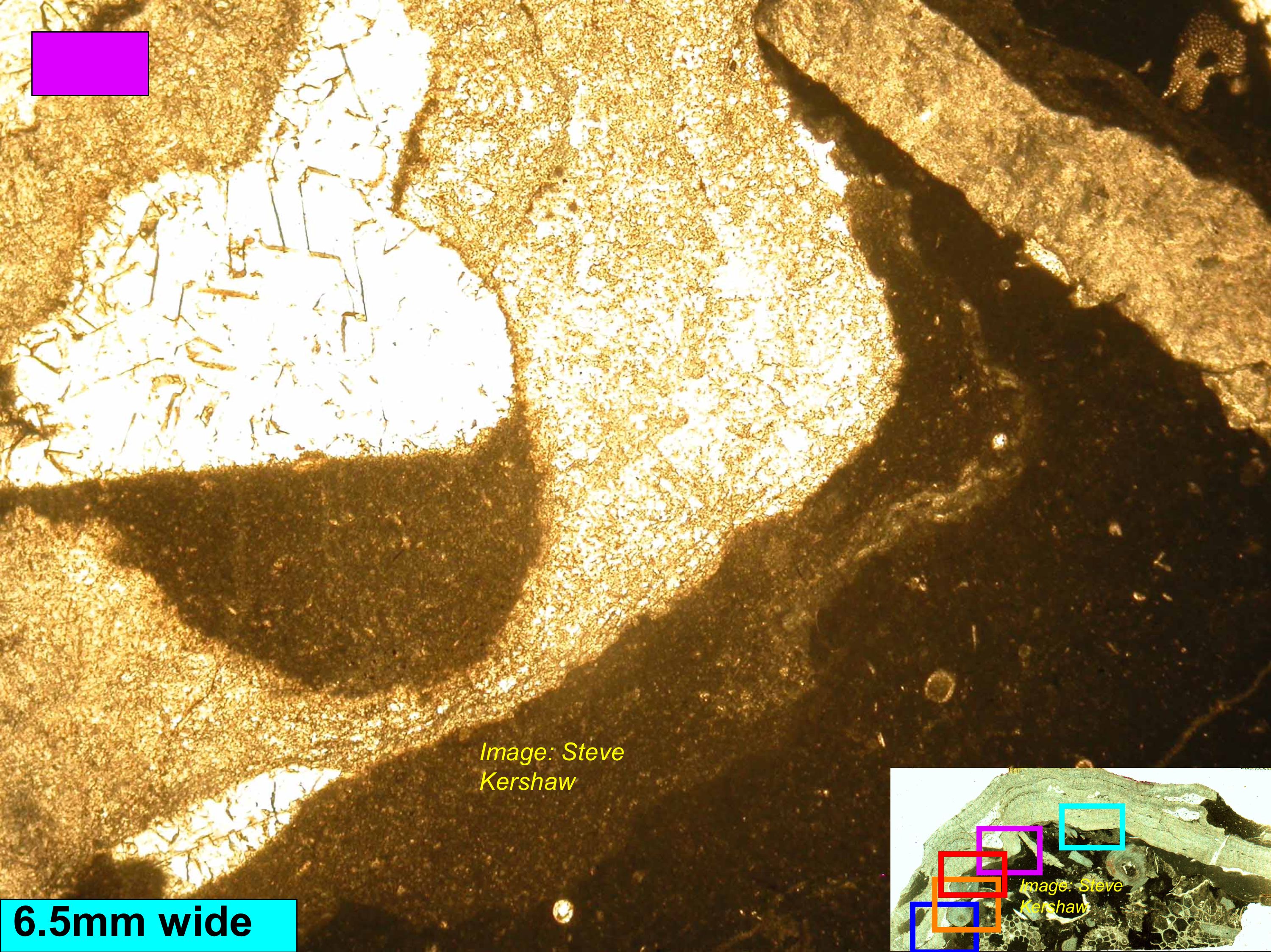
Figure 3A.20.

Figure 3A.21.
3B) EXAMPLES OF STROMATOPOROID-SUBSTRATE, AND CORAL-SUBSTRATE, RELATIONSHIPS, continued.

Figure 3B.1. Vertical thin section of Petridiostroma simplex, which grew on a complex of previous coral skeletons. At the base is the overturned corallum of a tabulate, encrusted by two other corals, prior to stromatoporoid growth. Stromatoporoid therefore appears to have grown on a topographic high provided by these corals. There may be partial development of primary cavities. Figures 3B.2-5 show details of boxes 1-4.

Figure 3B.2. Box 1 of Figure 3B.1. Margin of overturned coral encrusted by dark micrite (presumed microbial micrite), forming a substrate over which the stromatoporoid draped, thus no evidence of primary cavities.
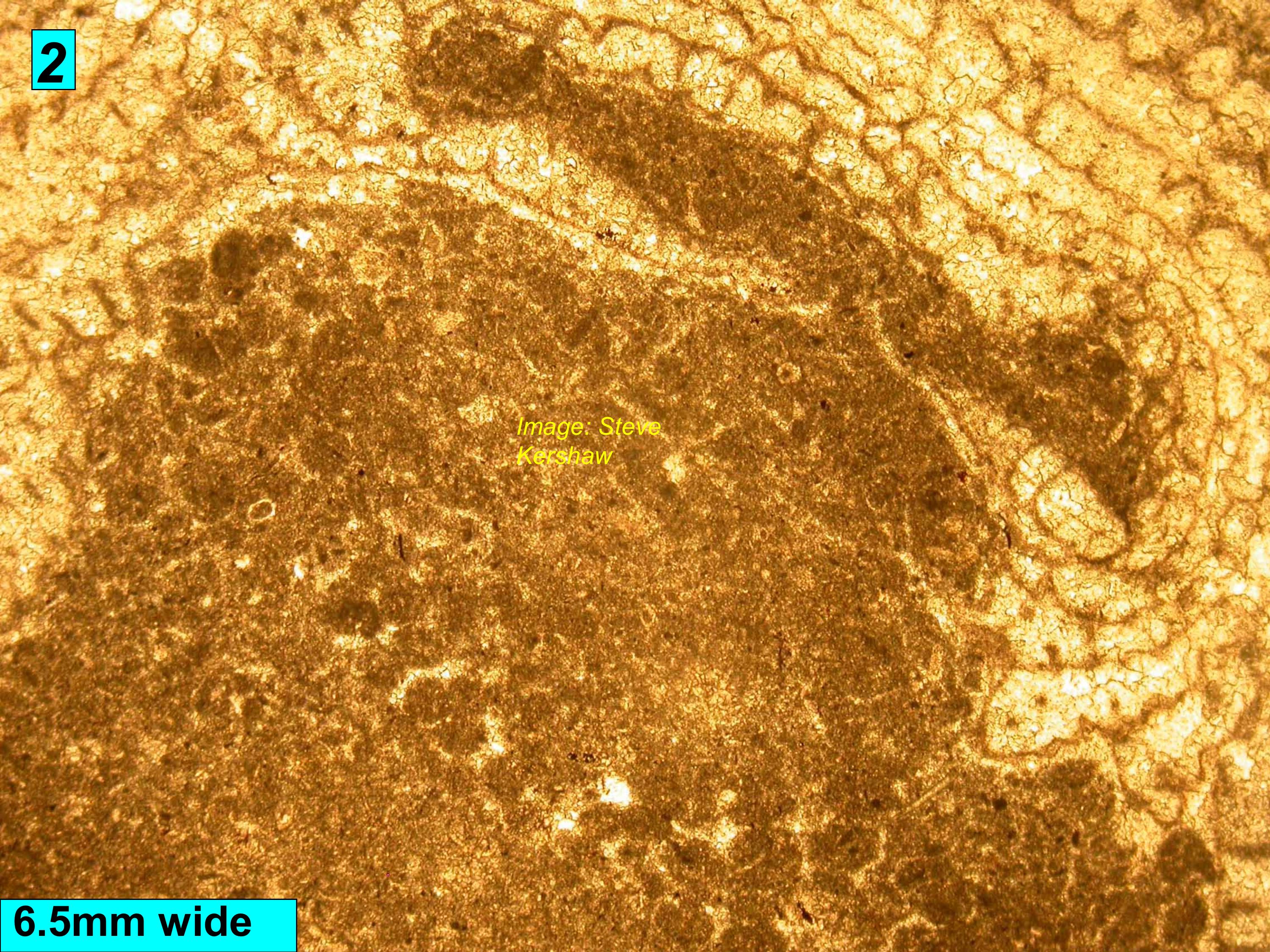
Figure 3B.3. Box 2 of Figure 3B.1. Stromatoporoid wrapped around a peculiar mass of micrite, that had topography and an indented margin, and was lithified on the sea floor, because the stromatoporoid laminae wrap around the sediment. It is possible this sediment and the corals formed a hardground upon which the stromatoporoid encrusted.


Figure 3B.4 & 5. Boxes 3 & 4 of Figure 3B.1.: Peculiar irregular topographic substrate highs encrusted by strom. It is presumed that the sediment was subsequently partly dissolved leaving voids, then infilled with cement; this presumption is because of the micro-irregular basal surface of the stromatoporoid.

Figure 3B.6. Vertical section through Densastroma pexisum, from Visby Formation, Lower Wenlock, Gotland, Sweden, showing details of ragged margin. Each interdigitation of stromatoporoid can be traced as a line into the interior of the strom, suggesting that these are growth interruption lines. However, the question remains: were the ragged margins 1) created by sedimentation followed by regrowth, or 2) did the stromatoporoid produce outward-growing flanges into open space, that were later backfilled? The second option seems more likely, since the first would require the stromatoporoid margin to grow in close contact with the sediment. Figures 3B.7 & 8 show enlargements.

Figure 3B.7.

Figure 3B.8.

Figure 3B.9. Densastroma pexisum growing on Eostromatopora impexa, with a thin layer of sediment between them, suggesting that the D. pexisum grew on the sediment on a topographic high provided by the dead E. impexa (that may have been killed by sedimentation). Englargements of boxes in next two slides.

Figure 3B.10. Detail of upper box in Figure 3B.9., showing a layer of sediment that matches the curve of the stromatoporoid base; did the stromatoporoid form a small primary cavity, just above the substrate?

Figure 3B.11. Detail of lower box in Figure 3B.9., showing a layer of sediment encrusted by a calcimicrobe, then more sediment, upon which the stromatoporoid sits. The calcimicrobe presumably encrusted firm or lithified sediment, and there may have been a primary cavity below the stromatoporoid.

Figure 3B.12. Vertical thin section of Densastroma pexisum showing an atrypid brachiopod (brachidium in section). Perhaps the brachiopod was deposited on the lower part of the stromatoporoid, then the upper part grew over it (and the sediment deposited along with the atrypid. There are other complexities in the lower part of this sample: what do you think they show? Enlargement of box in next slide.

Figure 3B.13. Enlargement of box in Figure 3B.12; note the stromatoporoid directly encrusts the brachiopod, suggesting the brach was deposited dead, and was overgrown by the strom, using its shell as substrate. Note the atrypid brachidium in section.

Figure 3B.14. Vertical thin section of Densastroma pexisum overgrown onto Pachystroma hesslandi. Inside the D. pexisum is an encrusting bryozoan, but this is apparently encrusting the sediment, and being overgrown by the stromatoporoid, not the more usual other way round. The next three photos are progressive enlargements that demonstrate the stromatoporoid is overgrowing the bryozoan.

Figure 3B.15.

Figure 3B.16.

Figure 3B.17.

Figure 3B.18. Vertical section through Densastroma pexisum, showing growth on hard substrate of a shell, that was subsequently partly dissolved. The presence of a soft-sediment burrow indicates the sediment was not firm when deposited, and the stromatoporoid is likely to have taken advantage of the hard surface of the shell, as a slight topographic rise on the substrate.

Figure 3B.19. Vertical section through Petridiostroma simplex, showing growth on sediment that was deposited on a rugose coral. The irregular shape of the sediment surface directly beneath the stromatoporoid once again suggests the stromatoporoid was growing on a solid surface and that the sediment had at least partly lithified before the stromatoporoid grew. Thus there is no clear indication of formation of primary cavities here.
3C) EXAMPLES OF STROMATOPOROID-SUBSTRATE, AND CORAL-SUBSTRATE, RELATIONSHIPS, continued.
TABULATE CORAL, WREN'S NEST, DUDLEY, ENGLAND
This coral was collected by Steve Kershaw in 1977 on a day visit, and it contains a range of interesting information about coral-substrate relationships, for comparisons with stromatoporoids. Only half the coral was collected; the other half is probably still there somewhere!

Figure 3C.1. Basal view of coral, showing the curved groove where the nautiloid lay on the sea floor. Although the coral began on the nautiloid, it extended beyond it onto the sediment, to the left of growth origin in this photo. The coral also shows growth rings on the base, a feature described in the section on Basal Lines later on this page.

Figure 3C.2. Vertical section of coral, showing growth banding, growth interruption and the difference between growth on the nautiloid and growth on muddy sea floor next to the nautiloid. It is obvious the coral grew more easily on the nautiloid shell.
Figures 3C.3-5 below show an acetate peel of the vertical section shown in Figure 3C.2, allowing more detail to be seen. The peel clearly shows the complexity of corallites on the sediment, while on the nautiloid the corallites propagated down the length of the nautiloid much more easily.
The peel also shows the reaction of the coral to a sedimentation event. The coral was partially killed by sediment, evident from the presence of sediment in the corallite tops in all three peel photos.

Figure 3C.3.

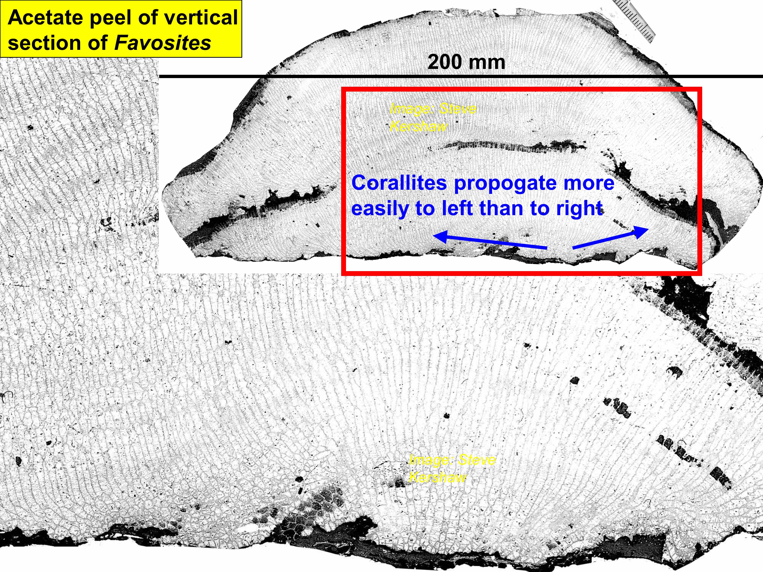
Figure 3C.4.

Figure 3C.5.
3D) EXAMPLES OF STROMATOPOROID-SUBSTRATE, AND CORAL-SUBSTRATE, RELATIONSHIPS, continued.
FRAMES WITH INTERPRETED PRIMARY CAVITIES

Figure 3D.1. Vertical section through a digitocolumnar-shaped stromatoporoid composed of vertical columns and lateral flanges that merge between columns, forming bridges. Beneath each bridge is a small area of sparite cement interpreted as void-filling cement in primary cavities. Thus the bridges formed in open space above substrate, linking columns together. Thanks to Nigel Watts, who gave the sample to Steve Kershaw.

Figure 3D.2. Enlargement of Figure 3D.1, showing details of bridges and void-filling cement.

Figure 3D.3. Framestone from reef core of Wenlock patch reef in Coates Quarry, Wenlock Edge, England. The frame is constructed by stromatoporoid genus Labechia, and contains some small areas of sparite interpreted as primary cavities (arrowed in photo below).

Figure 3D.4. Enlargement of Figure 3D.3., showing details of interpreted primary cavities.
Figure 3D.5-8 below show field photos of stromatoporoids and corals from the Llandovery Chicotte Formation on the southeastern coast of Anticosti Island, eastern Canada. All four photos show sparite cement beneath the fossils, interpreted here as primary cavities. Thanks to André Desrochers and Bill Ausich for field companionship in 2007 when these photos were taken.

Figure 3D.5.

Figure 3D.6.

Figure 3D.7.

Figure 3D.8.

Figure 3D.9. A small reef composed of only Petridiostroma linnarssoni, Visby Formation, Wenlock at Kneippbyn locality, Gotland, Sweden. This reef was figured in the famous old book on Gotland by Arie Manten in 1971, and subsequently by Paul Copper in 1983, celebrated as the 'smallest Upper Visby reef'. I collected several samples from this reef in 1976 and thin sections revealed that the reef is essentially a single, or perhaps multiple, skeleton(s) of one species of stromatoporoid, P. linnarssoni. The photos shown here demonstrate the presence of basal encrusting bryozoans on the base of the strom, and since the reef is in place, then either the encruster took advantage of a primary cavity, or alternatively later currents washed a cavity below the stromatoporoid used by the encruster. It is not possible to distinguish these two alternative explanations in this sample, but adds at least circumstantial evidence for primary cavities.
3E) EXAMPLES OF STROMATOPOROID-SUBSTRATE, AND CORAL-SUBSTRATE, RELATIONSHIPS, continued.
STROMATOPOROID AND CORAL BASAL LINES
|
Basal surfaces of corals and stromatoporoids often have concentric, or approximately concentric, lines, more accurately they are ridges. Ridges are often asymmetric, forming flanges inclined slightly downwards and facing away from the growth centre. Such flanges may be interpreted as caused by sedimentation killing the marginal portions of the skeleton, followed by regrowth. >>
|
However, many ridges are more-or-less symmetrical and not flange-form, and cannot be interpreted as caused by marginal sedimentation; the possibility that these are the roofs of primary cavities is enhanced by presence of similar features in some modern corals, which are primary cavity roofs.
Photographs in this section show examples of basal lines and considers their individual interpretation.
|

Figure 3E.1. Basal and vertical cross section views of Densastroma pexisum, Upper Visby Formation, Lower Wenlock, Gotland, Sweden. Prominent basal concentric rings are revealed in vertical section as sediment in marginal portions of successive stages of growth. Basal rings therefore indicate sedimentation interruption of marginal growth. Note that this stromatoporoid began growth on an orthoconic nautiloid.

Figure 3E.2. Basal and vertical section of a low profile stromatoporoid, Visby Formation, Lower Wenlock, Gotland, Sweden. Basal rings are abundant but small, with no obvious sedimentation-stimulated flanges.

Figure 3E.3 and 4. Basal view and oblique basal-vertical section view of another stromatoporoid with ridges but no sedimentation influence.

Figure 3E.4.

Figure 3E.5. Other samples lack the basal rings, even in the same species. These two specimens, viewed from side and basal views, are of Densastroma pexisum. Encrusters are present on the stromatoporoid bases. Note that the fact that the base is clearly visible is due to the relative ease with which sediment can be removed from the base, being clay-rich. Thus there is no apparent skeletal material on which the stromatoporoid grew, and thus the substrate appears to have been muddy. However, the presence of encrusters demonstrates existence of cavities at some stage in the development of the stroms. Thus it is unclear as to whether the stroms: A) grew on muddy sediment and were subsequently moved and deposited on an uneven substrate, allowing small cavities for encrusters to develop; or B) primary cavities formed but without basal lines. A third possibility, that the stromatoporoid was overturned, exposing the base, is less likely, because borers are not present on the basal surfaces of these samples. Kershaw (1980, in Lethaia) showed that borers occur only on upper surfaces of stroms unless they were overturned.

Figure 3E.6. Favositid tabulate, with growth lines, Visby Formation, Lower Wenlock, Gotland, Sweden. Note the lines in the central part are not apparently influenced by sedimentation, but in the outer parts sedimentation appears to have affected the lateral-spreading growth of the coral.

Figure 3E.7. Basal lines, encrusters and borers on base of Densastroma pexisum, and a step in the base of the stromatoporoid. Borers suggest the stromatoporoid was overturned, also therefore allowing for encruster growth. However, the basal lines are less easy to explain, possibly due to primary cavity growth, thus some or all of the encrusters may have attached in cavities.

Figure 3E.8. Enlargement of Figure 3E.7. Borers (B) and Encrusters (E).

Figure 3E.9. LEFT PHOTOS (Black and white): basal lines on a stromatoporoid (Petridiostroma linnarssoni) known to have grown in place. Note encrusting bryozoan. RIGHT PHOTOS: Basal lines on a sample of Densastroma pexisum that had been overturned and regrown. Note encrusting bryozoan. In these cases: 1) were the basal lines formed as a result of growth to form a primary cavity, or: 2) did the stromatoporoid grow directly on sediment, and the sediment subsequently removed by currents, allowing encrusters to grow?

Figure 3E.10. Basal and side views of modern scleractinian coral Diploria, showing basal lines, and a range of encrusters, particularly the noticeable Homotrema, red-coloured blobs. The central area of the coral shows breakage, because that was its attachment area to the substrate. Thus this coral formed a large primary cavity exposing most of its base for cryptic encrustation. Does the fact that it has concentric lines mean that the fossil examples in this webpage also grew with primary cavities?

Figure 3E.11. Basal view of a huge specimen of Labechia, which was famously sitting for many years by the gate of the original Allekvia Tingshus field station on Gotland. Apparently it is now in University of Lund. The Labechia is a low profile form, and shows a common characteristic of stromatoporoids, of merging of neighbouring specimens of the same species, indicating they were all alive together and presumably represent a series of sponges that settled in the same area of substrate. This one is unusual because of the spiral growth lines on the base, as mentioned in the text below the photo.
3F) CONCLUSIONS
The abstract of the paper by Kershaw et al. (2006), is given below. Please read this, then the subsequent text.
Reference: Kershaw, S., Wood, R. & Guo, L., 2006: Stromatoporoid response to muddy substrates in Silurian limestones. GFF, Vol. 128 (Pt. 2, June), pp. 131–138. Stockholm. ISSN 1103-5897.
|
Abstract: Stromatoporoids grew on both soft and hard substrates, and recent work revealed primary cavities in Devonian reef systems. We present evidence that stromatoporoids also developed primary cavities in muddy sediments. Stromatoporoids initiated on two categories of material: (1) fine-grained sediment, usually covering large bioclasts; (2) uncovered bioclasts, forming a small area of clean substrate. In both,subsequent growth was previously usually interpreted to be across, and in direct contact with, adjacent fine-grained sediment, itself presumed to be unlithified in most cases. However, stromatoporoid basal surfaces show two types of morphology: (A) smooth, generally curved and flat, and (B) corrugated, with concentric growth rings as minor ridges projecting downwards. Both may be present in different stromatoporoid individuals of the same species. Smooth bases are interpreted in most cases to indicate growth on unlithified sediment, and are common in category 1 above. >>
|
For corrugated bases, rings are interpreted to result from growth to form minor primary cavities by the stromatoporoid skeleton growing laterally a few mm above the substrate. The base then likely settled into the soft sediment as skeletal mass increased. Primary cavities are interpreted to be similar to those in muddy Devonian facies, but both differ from those in Devonian reefs (e.g. Canning Basin) where cavities may be tens of cm high, and were permanent. In the Silurian samples, a third morphology of concentric basal ring arrangement containing sediment wedges is interpreted as due to episodic sedimentation. Also, stromatoporoids grew on lithified and eroded intraclasts, indicating patchy early lithification of the sea floor in these open shelf settings. Evidence presented from the Silurian examples emphasises the dynamic response of stromatoporoids to their substrate, consistent with sediment-avoiding habits of modern calcified sponges.
|
|
The contents of these webpages show that the interpretations made by Kershaw et al. (2006), stated in the above abstract, are still current.
However, there is an additional point: the highly irregular basal portions of some stromatoporoids (particularly well illustrated in Figures 3A.8 & 9) may be interpreted to represent a partially or completely lithified sediment upon which the stromatoporoid encrusted. It is difficult to explain the irregular arrangements of laminae in this and other samples as primary cavity formation, since primary cavities in other stromatoporoids, illustrated here and by Rachel Wood's photos in Kershaw et al. (2006) show gently curved bases. Furthermore, it is difficult to accept that the stromatoporoid basal laminae may have physically pushed surface sediment into irregular forms as the sponge grew, not least because of the sediment aversion that modern sponges show.
Interpretation of irregular bases is less clear for tabulate corals, such as represented in Figures 3C.3 & 5; in these photos it is easier to accept that corallite propogation may have developed an irregular base by growth and budding of corallites, which was backfilled. >>
|
If sediment was lithified prior to stromatoporoid and coral growth in some cases, then this indicates hardgrounds in the Visby Formation. I have seen evidence of Trypanites borings in samples of limestone from the Visby Formation, but have no photos or samples to demonstrate here. Nevertheless, hardground formation in open marine shelf waters of the kinds of depths interpreted for the Visby (several tens of metres) suggests occurrence of synsedimentary lithification in these Silurian rocks to a greater extent than has been previously recognized.
Nevertheless, the occurrence of stromatoporoids with smooth bases, and of both stromatoporoids and corals from which the loose sediment can be removed to reveal the entire base, suggests an ability to grow on unconsolidated sediment; consequently the formation of primary cavities is equivocal in most specimens, rarely can it be proved. If stromatoporoids and tabulates were able to grow on the soft unconsolidated sediment directly, then the previous interpretations by myself (Kershaw 1998, published in Palaeontology), that stromatoporoid success in the Palaeozoic may be at least partly attributable to an ability to deal with soft substrates, remains a valid interpretation until someone proves it is wrong!
|
Figure 3F.10 below summarises the revised categories of substrate response in stromatoporoids.

Figure 3F.10. Schematic vertical section through a stromatoporoid showing the revised categories of substrate response. This diagram has not been previously published.
Click here to return to the GEOSCIENCE RESEARCH PROJECTS header page
Click here to return to the HOMEPAGE